The field of ophthalmology has had significant progress in the past few years, especially in the techniques that are employed in eye exams. These developments have improved diagnosis accuracy, but they have also completely changed patient expertise, making eye exams more effective, comfortable, and informative than they have ever been. If you are looking for Comprehensive Eye Exams in Brampton, feel free to contact Drs J & K Gill & Associates. As seasoned experts, they are aware of the need to have the appropriate equipment for diagnosing, evaluating, and tracking ocular disorders. Further, underlying medical concerns such as diabetes, thyroid disorders, cancers, autoimmune illnesses, and hypertension can all be found with a comprehensive eye exam. This article explores the newest technology influencing eye exams today and discusses their advantages and contributions to eye health.
Digital Scanning of the Retinal Layer
The way eye care specialists record and keep track of ocular health has changed dramatically with the advent of digital retinal imaging. This technology uses specialized cameras to take high-definition pictures of the retina. These pictures give a thorough picture of the retina’s health, making it possible to identify anomalies like tumors, retinal detachment, and indications of systemic illnesses like high blood pressure and diabetes. By providing a visual representation of any issues or changes in ocular health over time, digital retinal imaging also makes interacting with patients and eye care specialists easier.
Tomography of Optical Coherence (OCT)
Optical Coherence Tomography (OCT) is among the most significant developments in the field of eye care diagnosis. With this non-invasive imaging method, cross-sectional images of the retina with great resolution are obtained using light waves. Early detection and monitoring of many eye disorders, including macular degeneration, diabetic retinopathy, and glaucoma, are made possible by OCT, which provides detailed images of the interior tissues of the eye, including the layers of the retina, optic nerve, and macula. OCT also helps in monitoring the course of diseases and determining how well therapies work, which eventually results in improved ocular health management.
The Topography of the Cornea
With the aid of a diagnostic technique called corneal topography, one can accurately assess the thickness and curvature of the cornea by mapping its surface in detail. Astigmatism, keratoconus, and imperfections in the cornea can all be diagnosed and treated with the use of this technology. Eye care specialists can create personalized treatment plans for refractive operations like PRK or LASIK by analyzing the topographical data, guaranteeing the best possible results for their patients. By giving precise measurements for lens design and selection, corneal topography also helps with contact lens fitting, especially for patients with uneven corneas.
Automation Refraction Devices
In the past, the optometrist or ophthalmologist would manually adjust the patient’s glasses based on their subjective feedback. However, by precisely measuring a patient’s refractive error using complex algorithms and digital imagery, automated refraction systems have completely changed this procedure. In addition to giving more precise prescriptions for glasses or contact lenses, these technologies expedite the examination procedure, saving each patient time and avoiding errors related to manual refractive procedures.
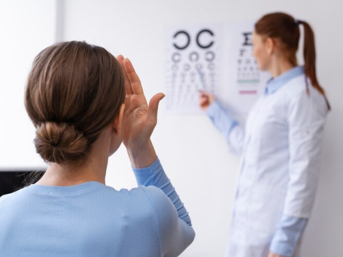
Examining the Visual Field
Assessing peripheral vision and identifying any anomalies or loss of vision that can point to underlying neurological disorders like brain tumors or ocular diseases like glaucoma need the use of visual field tests. With the use of complex algorithms and cutting-edge stimulus presentation methods, contemporary visual field testing technology can accurately assess a patient’s sensitivity to visual fields and identify minute changes over time. To preserve vision and enable prompt intervention in the event of progressive eye disease, these tests are essential for early detection and monitoring of the condition.
Last Thoughts
Modern eye exams now include state-of-the-art technology that greatly improves the accuracy, effectiveness, and thoroughness of ocular diagnostics. These advancements enable eye care providers to deliver better patient care by facilitating early diagnosis and treatment of eye diseases. Examples of these advances include automated refraction devices, corneal topography, and advanced imaging techniques like OCT and digital retinal imaging. Future developments in the field of eye care appear promising as long as technology keeps developing; these developments will eventually help patients all over the world maintain and improve their ocular health and eyesight. To learn more about modern eye exams, you can go through the Drs J & K Gill & Associates. They provide their expert guidance with Comprehensive Eye Exams in Mississauga and its nearby areas.